


Hey there! I'm a supplier of 10 - lead EKG leadwires, and today I wanna chat about what the purpose of each lead in these 10 - lead EKG leadwires is. Now, EKG, or electrocardiogram, is a super important tool in the medical field. It helps doctors get a peek at the electrical activity of the heart, and the leadwires are what make that possible.
Let's start by understanding the basics. EKG leadwires are like the messengers that carry the electrical signals from the patient's body to the EKG machine. Each lead has a specific job, and together, they give us a comprehensive view of the heart's function.
The Limb Leads
The first set of leads we'll talk about are the limb leads. There are four of them: right arm (RA), left arm (LA), right leg (RL), and left leg (LL). These leads are placed on the limbs, as the name suggests.
The RA lead is placed on the right wrist or the right shoulder area. Its main purpose is to pick up the electrical signals coming from the right side of the heart. It helps in detecting any abnormalities in the right atrium or the right ventricle.
The LA lead, on the other hand, is placed on the left wrist or the left shoulder. It focuses on the electrical activity on the left side of the heart. This lead is crucial for spotting issues in the left atrium and the left ventricle.
The RL lead is often used as a ground lead. It helps to reduce electrical interference and provides a stable reference point for the other leads. It's placed on the right ankle or the right lower leg.
The LL lead is placed on the left ankle or the left lower leg. It works in conjunction with the other limb leads to create different views of the heart's electrical activity. The combination of these limb leads gives us what are called the bipolar and augmented unipolar leads.

The bipolar leads, which are formed by the RA - LA, RA - LL, and LA - LL combinations, give us a view of the heart from different angles. For example, the RA - LA lead shows the electrical activity between the right and left arms, providing a horizontal view of the heart.
The augmented unipolar leads, aVR, aVL, and aVF, are derived from the limb leads as well. The aVR lead gives us a view from the right shoulder, looking down at the heart. The aVL lead looks at the heart from the left shoulder, and the aVF lead views the heart from the feet. These augmented leads help in detecting specific types of heart problems, like arrhythmias or myocardial infarctions.
The Chest Leads
Now, let's move on to the chest leads. There are six of them, labeled V1 through V6. These leads are placed on the chest, in specific positions over the heart.
The V1 lead is placed in the fourth intercostal space on the right side of the sternum. It provides a view of the electrical activity in the right ventricle. This lead is great for detecting right - sided heart issues such as right ventricular hypertrophy.
The V2 lead is placed in the fourth intercostal space on the left side of the sternum. It also focuses on the right ventricle but provides a slightly different perspective compared to V1.
The V3 lead is placed between V2 and V4. It gives us a view of the septum, which is the wall that separates the left and right ventricles. Abnormalities in the septum can be detected using this lead.
The V4 lead is placed in the fifth intercostal space at the mid - clavicular line. It mainly looks at the anterior part of the left ventricle. This lead is important for diagnosing anterior wall myocardial infarctions.
The V5 lead is placed between V4 and V6, at the anterior axillary line. It provides a view of the lateral part of the left ventricle.
The V6 lead is placed at the mid - axillary line in the fifth intercostal space. It focuses on the far - lateral part of the left ventricle.
The chest leads are really important because they give us a detailed view of the heart's electrical activity in the horizontal plane. They can help in detecting a wide range of heart conditions, from minor arrhythmias to major heart attacks.
Why 10 - Lead EKG Leadwires Matter
You might be wondering, why do we need 10 leads? Well, having 10 leads gives us a more complete picture of the heart's electrical activity compared to fewer leads. Each lead provides unique information, and when we combine all of them, we can detect heart problems that might go unnoticed with a smaller number of leads.
For example, a patient might have a small area of damage in the heart that only shows up on a specific lead. If we don't have that lead, we might miss the diagnosis. That's why 10 - lead EKG leadwires are so important in modern medicine.
Our Product Range
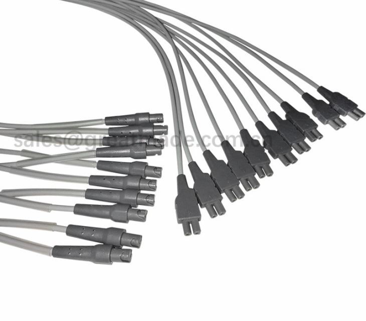
As a supplier of 10 - lead EKG leadwires, we offer a wide range of products to meet different needs. We have leadwires that are Compatible GE Universal Leadwire 2001925 - 003, which are designed to work seamlessly with GE EKG machines. These leadwires are made with high - quality materials to ensure accurate signal transmission and long - term durability.
We also have the Welch Allyn CT 100 10 - lead leadwires. These are specifically designed for Welch Allyn CT 100 EKG machines. They are easy to use and provide reliable results.
And if you're using an Esaote EKG machine, we have the EKG 10 lead for Esaote. These leadwires are a perfect fit for Esaote devices and are built to last.
Conclusion
In conclusion, each lead in a 10 - lead EKG leadwire system has a specific purpose. The limb leads give us a view of the heart from different angles in the vertical plane, while the chest leads provide a detailed view in the horizontal plane. Together, they help doctors diagnose a wide range of heart conditions accurately.
If you're in the market for high - quality 10 - lead EKG leadwires, we're here to help. Whether you need leadwires for a GE, Welch Allyn, or Esaote EKG machine, we have the right products for you. Don't hesitate to reach out if you have any questions or if you're interested in purchasing our leadwires. We're always happy to have a chat and discuss your needs.
References
- Goldberger, A. L., Goldberger, E. R., & Shvilkin, A. (2006). Clinical Electrocardiography: A Simplified Approach. Mosby.
- Fisch, C., & Knoebel, S. B. (2000). Electrocardiography of Clinical Arrhythmias. Futura Publishing Company.